Monday, August 31, 2009

Reduced BMD in adults with very low birth weight may lead to osteoporosis, related fractures


Orthopaedics Today

1st on the web (August 28, 2009)

Individuals who had a very low birth weight (VLBW) had significantly less bone mineral density (BMD) at the adult age when they should normally reach peak bone mass compared to same-aged adults born at term, Finnish researchers found.

In the Helsinki Study of Very Low Birth Weight Adults, a multidisciplinary cohort study designed to see if health problems in VLBW babies persist into adulthood, Petteri Hovi, MD, and colleagues evaluated 144 adults between 18.5 and 27.1 years old born in the greater Helsinki area between 1978 and 1985 with VLBW. They compared the group’s skeletal health to 139 adults matched for age, gender and birth hospital.

Hovi and colleagues defined VLBW babies as those whose birth weight was less than 3.31 pounds.


The researchers determined each participant’s BMD using dual-energy X-ray absorptiometry. They calculated Z scores representing the participants’ lower lumbar spine and hip BMD, defining reduced BMD as a Z score of -1.0 units or less.

The study group’s average Z score for the lower lumbar spine was -0.51 unit (95% CI: 0.28-0.75); the average femoral neck Z score was -0.56 unit (95% CI: 0.34-0.78) for areal BMD. After adjusting for the shorter adult height and less intense level of exercise practiced in the VLBW group, the differences remained statistically significant, according to a press release.

“Furthermore, 44% of the VLBW participants, but only 26% of the term-born participants, had a lumbar spine Z score of -1.0 or less,” study editor Tom W.J. Huizinga, of Leiden, Netherlands, wrote in his summary of the findings.

Hovi and colleagues said in their abstract, “This finding may predict symptomatic osteoporosis and increased fracture rates.”

In addition, increased vigilance in osteoporosis prevention may be warranted in VLBW children who become adults with low BMD, they noted.

Researchers said the main limitations of the study were the final cohort may not be representative of the original cohort of individuals born in Helsinki-area hospitals; comparisons they made were only internal; and they measured BMD just once, which reduced the chances of associating lower BMD with other possible causes, such as delayed skeletal maturing in the VLBW group.

Reference:

  • Hovi P, Andersson S. Järvenpää A-L, et al. Decreased bone mineral density in adults born with very low birth weight: A cohort study. PloS Med. 2009;6(8):e1000135. DOI: 10.1371/journal.pmed.1000135.

Orthopedic study suggests knee extensor strength plays a part in keeping osteoarthritis at bay

Orthopaedics Today

1st on the web (August 27, 2009)

Researchers exploring factors that affect the risk of developing symptomatic knee osteoarthritis in a multicenter study found that women with the strongest thigh muscles developed the condition less often than women with weaker knee extensor strength.

According to findings from the Multicenter Knee Osteoarthritis Study, funded by the U.S. National Institute on Aging, men with stronger thigh muscles were only slightly more protected from developing symptomatic OA than those with weaker muscles. Furthermore, thigh muscle strength was not helpful for predicting radiographic OA in women or men, the investigators noted in their study, which appears online today in Arthritis Care & Research.

“These findings suggest that targeted interventions to reduce risk for symptomatic knee OA could be directed toward increasing knee extensor strength,” co-investigator Neal A. Segal, MD, MS, from the department of orthopedics and rehabilitation at the University of Iowa, said in a press release.


Segal and colleagues studied the knees of 3,026 men and women 50 to 79 years old. They measured study participants’ thigh muscle strength with a dynamometer and determined their H:Q ratio, an indication of the balance of strength between hamstrings and quadriceps.

Investigators followed subjects 30 months using knee radiographs and telephone interviews to establish baseline OA, pain and other knee symptoms with the goal of determining whether knee extensor strength was a predictor of radiographic or symptomatic knee OA.

To draw their conclusions, they took into account the participants’ height, weight, body mass index, femoral neck bone mineral density and self-assessed level of physical activity.

At the end of their study, investigators reported that 7.1 % of men and 9.9% of women developed radiographic knee OA, compared to 7.8% of men and 10.1% of women who had signs of symptomatic OA. “H:Q ratio was not predictive of symptomatic knee OA in either men or women,” they wrote in their results.

The lack of hip abductor strength assessments in the analysis was one of the study limitations, the researchers noted in the press release.

“Study of hip abductor strength, which is important for control of the knee joint, may be useful in a more comprehensive study of risk for OA of the knee,” Segal said in the release.

Reference:

  • Segal NA, Torner JC, Felson D, et al. Effect of thigh strength on incident radiographic and symptomatic knee osteoarthritis in a longitudinal cohort. Arthritis Care Res. Published online Aug. 27, 2009 (DOI 10.1002/art).

Tuesday, August 25, 2009

Limb-sparing surgery and amputation provide similar quality of life for patients with bone cancer


1st on the web (August 11, 2009)

Limb-sparing surgery, an alternative to amputation for bone and soft tissue sarcomas of the lower limb, may not provide much or even any additional benefit to patients compared to amputation, according to a new review.

Researchers who conducted the analysis, which was posted yesterday in the online edition of Cancer, recommend that patients and physicians should rethink the pros and cons of both limb-sparing surgery and amputation before making a final decision.

To compare the costs and benefits of limb-sparing surgery with amputation in these patients with cancer, researchers Ronald Barr, MD, MB, ChB, of McMaster University in Hamilton, Ontario, and Jay Wunder, MD, MSc, of Mount Sinai Hospital and the University of Toronto, reviewed all published papers on limb-sparing surgery that also measured patients’ functional health and quality of life.


advertisement

According to a press release, the researchers found that while limb-sparing surgery is generally as effective as amputation in removing the patient’s cancer, it tends to be associated with more early and late complications. Studies also show that, particularly for patients with lower limb bone sarcomas, limb salvage does not provide a better quality of life to patients than amputation, according to the press release.

In the press release, Wunder said most studies have found that the differences in disability between amputation and limb-sparing patients are smaller than expected. Many revealed no significant differences in psychological health and quality of life between patients who underwent amputations and those who had limb-sparing surgery. However, there appear to be greater advantages to limb-sparing surgery over amputation for higher surgical sites in the lower limb, such as the hip.

Some studies have compared the costs of amputation vs. limb-sparing surgery. “Up front” surgical costs, the duration of rehabilitation and the need for revisions are all greater for limb-sparing surgery. However, amputation carries greater longer-term costs related to artificial limb manufacture, maintenance and replacement, the researchers said in the press release.

The authors say additional research is needed to provide a thorough comparison of amputation and limb-sparing surgery in different types of patients with bone and soft tissue sarcomas.

“Future studies that include function, health-related quality of life, economics and stratification of patients by age will be useful contributions to decision-making … by patients, health care providers and administrators,” Wunder said in the press release.

Reference:

  • Barr R, Wunder J. Bone and soft tissue sarcomas are often curable – But at what cost? A call to arms (and legs). Cancer. Advance preview published on Aug. 10. 10.1002/cncr.24458.

Promising results seen with minimally invasive repair of Achilles tendon ruptures


Surgeon cautions that device may not benefit tendon rupture cases with frayed ends.

By Gina Brockenbrough
ORTHOPEDICS TODAY 2009; 29:42

DENVER — With minimally invasive surgical repair of Achilles tendon ruptures, surgeons look to obtain the advantages of open repair techniques while avoiding the problems of wound complications and infections.

At the American Orthopaedic Foot and Ankle Society’s 24th Annual Meeting, Eric M. Bluman, MD, PhD, discussed the results of minimally invasive repair techniques and highlighted the outcomes of repairs using a minimally invasive Achilles tendon suture device (Achillon, Integra).

“I think that minimally invasive repairs have shown promising results in case series and limited level I studies,” Bluman said. “Obviously, more level I studies are needed, and I think that we need to show caution in using [the Achillon] technique. It may be particularly difficult to use in ruptures in which the tendon ends are frayed or in very distal ruptures where tendon material in which to put your sutures in is limited.”


advertisement
Cipka

Tendon suture device

Although his presentation focused on minimally invasive repairs, Bluman use nonoperative treatment for Achilles tendon ruptures when indicated and open repair on occasion. Regarding surgical methods, “I began questioning how I could justify the assault on the posterior tissues in the ankle, when there are methods to obtain repairs with much less invasive methods,” he said.

The minimally invasive repair technique he uses is aided by the Achillon device. The technique uses a 2-cm vertical incision that allows for direct visualization of tendon apposition. It also maintains all the sutures deep to the peritenon, Bluman said. “One of the nice things about the vertical incisions is it is easily converted into an open approach if needed.”

Achilles tendon
Image showing the scar left after a minimally invasive Achilles tendon rupture repair.

Image: Bluman EM

Bluman said he has used the technique on many patients, 11 of whom have a minimum 1-year follow-up. “All are back to their pre-rupture activity level,” he said. “These are active duty military people and active retirees. All patients are satisfied and would have the procedure again.”

Comparing the uninjured side to the contralateral limb using dynamometry showed no significant difference, he said. He noted no wound problems, infections, re-ruptures, sural nerve injuries or adhesions were seen.

No infections, nerve injuries

A study by the device’s inventor reported on 83 patients whose tendons were repaired using the Achillon device showed no infections and no sural nerve injuries, Bluman said. “They did have three re-ruptures which they attributed to noncompliance of the patients, and all of their patients did eventually return to previous function.”

He also cited an independent study performed by Calder, and colleagues which corroborated the results with the device in 25 patients. The investigators found no infections, wound dehiscence, sural nerve injuries or re-ruptures and reported good functional results.

Although totally percutaneous techniques reduce infection and wound dehiscence rates to 0%, “There have been reports of increased rates of sural nerve injury as high as 17% in some cases,” Bluman said. “There is an inability to directly visualize tendon apposition as well.” Sural nerve injuries have been shown to be due to lassoing of the nerve with the suture in percutaneous cases.

In a study comparing percutaneous and open repair, Lim and colleagues found two adhesions, two re-ruptures and a 21% infection rate in the open repair group. In the percutaneous cohort, they had one re-rupture, one sural neuropathy and had three cases of wound puckering. “This study was limited in that no dynamometry was done and only subjective outcome measures were used for evaluation,” Bluman said.

For more information:
  • Eric M. Bluman, MD, PhD, can be reached at Madigan Army Medical Center, Orthopaedic Surgery, 9040 A. Fitzsimmons Fr., Tacoma, WA 98431; 253-968-1581; e-mail: emb43@cornell.edu. He receives miscellaneous non-income support from DePuy.
Reference:
  • Bluman EM. Minimally invasive surgical repair of the Achilles tendon rupture. Presented at the American Orthopaedic Foot and Ankle Society 24th Annual Summer Meeting. June 26-28, 2008. Denver.

Total Hip Replacement and Hip Resurfacing Surgery

Source: http://www.bananarepublican.info/Hip_Surface_Replacement.htm

Background

Hip replacement surgery has been around since the early 1960s. Sir John Charnley experimented in the early 1950s, and he used a small (22 mm) stainless steel ball on a stem in 1962 that he inserted into the femur (hip) bone to replace the femoral head (ball). He then inserted a high-density plastic socket to replace the acetabular (socket) side of the hip joint. Both were secured with a self-curing acrylic polymer known as bone cement.

Total Hip Replacements

Today, the modular balls are made of a cobalt-chrome metal alloy or a ceramic material, and some of the components are press-fit and do not require bone cement. The procedure remains basically the same: (1) the femur bone is amputated to remove the femoral head; (2) the femoral canal is reamed-out for insertion of the stem; (3) an acetabular socket is affixed to the socket side of the hip; and (4) the ball joint is inserted into the acetabular socket. This is known as a total hip replacement, or more correctly, total hip arthroplasty (THA).

image from www.wmt.com

(Click on image to enlarge)

image from www.jri-oh.com

(Click on image to enlarge)

image from www.jri-oh.com

(Click on image to enlarge)

The acetabular socket used in THA is normally lined with a high molecular weight polyethylene (sometimes the liner is ceramic). A metal or ceramic ball is attached to the stem and rotates within the socket. Fine particulate debris is produced from the wearing process of the ball against the liner that leads to tissue reaction. The body’s immune system attacks the debris, and consequently, attacks the adjacent bone supporting the THA device, leading to bone loss and a loosening of the device. This bone loss is known as osteolysis. To lessen the amount of wear, a small ball (approximately 30 mm) is used; however, the small size of the ball makes the joint less stable and increases the risk of dislocation in certain circumstances.

The loosening of the THA device requires revision surgery in which a larger diameter stem must be inserted in the femoral canal. Depending on the age and activity of the patient, multiple revision surgeries may be necessary throughout a patient’s life. A young (under 60), active individual can expect only 10 – 15 years before needing revision surgery. Revision surgery can be complex and costly. The lifespan of a THA device is clocked in miles rather than years.

(Note: Wright Medical Technology, Inc. has developed a large femoral head using metal-on-metal technology (see Hip Surface Replacement below) that reduces the risk of dislocations and osteolysis in THRs. The large head THR has received FDA approval and is actively being marketed.)

Hip Surface Replacements

Although it was experimented with and attempted in the 1960s, metal-on-metal “resurfacing” of the femur and acetabulum was abandoned because of loosening of the fittings. With the refinement of acrylic fixation and its very successful use with the THA stem, interest in hip resurfacing was renewed, and it was subsequently used in several countries in the 1970s. (See History of Hip Resurfacing.)

Resurfacing has the advantage of preserving the femoral bone stock (and marrow contained in the femur). It also has the advantage of easy future revision to THA if it becomes necessary. Since the femur is persevered and not amputated in the initial hip surface replacement surgery, it is available to support a THA stem should revision become necessary. Maintaining the integrity of the femur bone also aids in the mechanical transfer of weight and stress in a more natural manner. Where THA patients often experience thigh pain, recipients of hip surface replacements avoid that particular discomfort.

image from www.jri-oh.com

(Click on image to enlarge)

image from www.jri-oh.com

(Click on image to enlarge)

Using a metal acetabular socket as well as a metal cap over the femur head (metal-on-metal) eliminates the polyethylene debris produced in THA. The metal wear debris from a hip surface replacement produces smaller particles than polyethylene wear debris. The inflammatory response to metal debris is considerably less than that from polyethylene debris. It is believed that the body can partially dissolve and expel metal since it is a naturally occurring substance in the body. There is concern by some of the toxicity of metal, but there is currently no definitive evidence that metal ions cause cancer. Since a metal surface does not wear as readily as a polyethylene lining, a larger ball (approximately 38-51 mm) can be used that adds stability to the joint and reduces the danger of dislocation.

images from www.wmt.com & www.jri-oh.com

(Click on images to enlarge)

The surgery time for hip surface replacement is slightly longer than that for THA. The attachment of the acetabular socket is basically the same. It is press-fitted and does not require bone cement. The attachment of the cobalt-chrome cap requires a more precise alignment, and it takes slightly longer to fit. The hole for the pin insertion must be aligned and drilled, and the dome of the femoral head must be ground and shaped to fit the cap. Some bone cement is used to affix the cap, but the interior surface of both the cap and the socket is such that bone grows into the relief surface to grip the device. (See the video clips of hip resurfacing surgery.)

The following images are from an Instructional Lecture delivered at an International Symposium in Fukuoka, Japan on March 16, 1996 by Harlan C. Amstutz, Peter Grigoris, and Frederick J. Dorey entitled "Evolution and future of surface replacement of the hip." Journal of Orthopaedic Science. J Orthop Sci (1998) 3:169-186.

Superimposed hemisurface. Pin centering guide. Cylindrical reamer.

Saw cutoff guide and oscillating saw. Chamfered reamer. Femoral head bone preparation.

(Click on images to enlarge)

Risks involved in the hip surface replacement surgery are the same as the risks involved in any major surgery. Risks specific to the hip surface replacement involve the potential for cracking in the neck of the femur bone due to the drilling of the guide hole through the neck for the support pin in the metal cap, and also a negative reaction of the femur head to dislocation and being reshaped to fit the metal cap leading to the development of avascular necrosis (bone death)--often referred to as AVN--due to a disruption of blood circulation to the femur head and neck (see AVN Risk). In such instances, a THA could easily be performed to correct the problem.

Hip surface replacement in the United States has been pioneered by Harlan C. Amstutz, M.D. at the Joint Replacement Institute in Los Angeles, CA. For years, a hip surface replacement in the United States has been labeled an “investigative device” by the Food and Drug Administration (FDA). The longest study has been conducted by Wright Medical Technology, Inc. under the product name of CONSERVE ® Plus Total Resurfacing Hip System. The clinical trials have proceeded for a number of years, and they are nearing their end. They have involved nine surgeons across the country in California, Florida, Texas, Maryland, North Carolina, Ohio, and in the Pacific Northwest. Corin Medical, Ltd. of the United Kingdom has also begun an FDA study in the United States using the Cormet 2000 device.

Click here to read the 2-6 year follow up report of the first 400 CONSERVE ® Plus hips.

In Europe, the Birmingham Hip Surface Replacement System (BHR) has been in use for many years. Smith & Nephew Inc., manufacturer of the BHR, applied for FDA approval, and perhaps due to the long record of use in Europe, they obtained FDA premarket approval to begin commercial distribution of their device in the United States on May 9, 2006 (see FDA approval letter and FDA announcement). Because the BHR was not previously used in the United States, the number of American surgeons qualified to use it was limited due to the fact that they had all been participating in the Wright and Corin studies; however, that is destined to change with the FDA approval obtained by Smith & Nephew.

Hospital for Special Surgery lays claim to being the oldest U.S. orthopedic hospital


The 162-bed hospital is known for achievements in sports medicine, joint replacement and research.

By Susan M. Rapp
ORTHOPEDICS TODAY 2009; 29:58

The 146-year history of Hospital for Special Surgery is rich with clinical, research and educational accomplishments that have improved how orthopedic medicine is practiced.

Pioneers

In 1863, Hospital for Special Surgery (HSS) in New York began as the Hospital for the Ruptured and Crippled in a philanthropic effort to provide medical care to injured Civil War soldiers and needy city residents. It has been led by 11 surgeons-in-chief and now has 85 full-time orthopedic surgeons who performed 24,000 orthopedic procedures in 2008, including more than 7,366 joint replacements, according to current HSS Surgeon-in-Chief Thomas P. Sculco, MD.

The list of musculoskeletal medicine pioneers from HSS reads like a who’s who of orthopedic innovation: Harlan C. Amstutz, MD, Albert Burstein, PhD, John N. Insall, MD, Chitranjan S. Ranawat, MD, Peter S. Walker, PhD, and Russell E. Warren, MD, among others.

“The many strong physician contributors at HSS over the years are typified by how well they integrated excellent patient care with critical review and analysis of clinical results and applied, basic science,” said Douglas W. Jackson, MD, Chief Medical Editor of Orthopedics Today.

Hospital for Special Surgery
Hospital for Special Surgery (HSS) is an independent, free-standing orthopedic hospital that is closely affiliated with a medical center and the Weill Cornell Medical College for conducting musculoskeletal research.

Images: Hospital for Special Surgery

Leading the way

“We are the oldest orthopedic hospital, certainly in the United States and probably in the world,” Sculco said.

HSS was founded by the first surgeon-in-chief, James Knight, MD, whose interest in orthopedics was limited to designing and constructing braces for children’s congenital deformities.

Eventually HSS’ focus shifted entirely to the treatment and rehabilitation of the musculoskeletal system for patients locally and around the world, which remains the mission of the 162-bed hospital. HSS also has

Surgical innovation

An early HSS pioneer, Virgil P. Gibney, MD, became the second surgeon-in-chief in 1887 and held the position 40 years. He instituted changes in the work performed at the hospital and wrote about hip surgery.

“Virgil Gibney was responsible for establishing the first orthopedic residency,” in the 1890s, Sculco told Orthopedics Today.

“He put the hospital on the map as a surgical hospital,” said David B. Levine, MD, Director of HSS Alumni Affairs.

James Knight, MD
The first HSS Surgeon-in-Chief James Knight, MD, founded the hospital in 1863 at his home in lower Manhattan. He was a general physician rather than an orthopedic surgeon.

John Marshall, MD
The late John Marshall, MD, a physician at HSS, is credited with launching sports medicine as an academic discipline.

Gibney’s leadership

Gibney was the first president of the American Orthopaedic Association.

Under Philip D. Wilson Sr., MD, who became the fifth surgeon-in-chief in 1935, the hospital changed its name to The Hospital for Special Surgery, moved to its current site and affiliated with Weill Cornell Medical College. In 1996, HSS dropped “The” from the beginning of its name.

“Wilson, Sr. had the vision to make it a very specialized institution and improve its research and academic mission,” Sculco said.

Wilson was AAOS president in 1934. His son, Philip D. Wilson Jr., MD, was the eighth surgeon-in-chief and AAOS president in 1972.

Knee arthroplasty

Wilson Jr. bridged the gap between engineering and biomechanics, according to Sculco.

“He created an environment here in the late 1960s and early 1970s for the design and development of implants with surgeons collaborating closely with engineers and basic scientists. To a large extent, that still goes on today.”

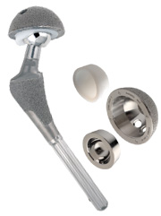
The joint arthroplasty research work performed by engineers like Walker and Burstein exemplifies that collaboration, yielding the duo-condylar knee developed in 1971. In 1973 Walker broached the concept of the cemented cruciate-sacrificing total condylar knee prosthesis that improved on other available designs.

Burstein and Insall developed the first posterior stabilized knee prosthesis in 1979 and in 1989 an updated version was marketed.

Subspecialization

In the 1970s, “[Wilson, Jr.] reorganized the orthopedic department into subspecialties and anatomic regions,” Levine said. “He was way ahead of his time.”

Specialty clinics for treating groups of patients with similar problems followed.

Sculco said this change at HSS mainly influenced what happened nationally and started the trend toward subspecialization. More importantly, Wilson, Jr. created an environment where individual doctors and researchers could flourish.

The research accomplishments at HSS are as strong as its clinical areas with work underway in tendon and ligament repair, soft tissue healing, osteolysis, spine, sports medicine, cartilage repair, osteoarthritis and other areas. The hospital has a strong commitment to registries and has several, including a prospective total joint replacement registry with data on nearly 13,000 patients and one containing more than 20,000 retrieved implants.

Brace shop
Workers at the HSS brace shop are shown working in the early 1900s. Bracing was a large component of treatments at the hospital. More recently, orthopedic implants were fabricated on-site at the hospital.

Retrieval program

Timothy Wright, PhD, Director of the Department of Biomechanics started the HSS retrieval program in 1977 with Burstein.

The research and biomechanics programs Walker had in place when Wright joined HSS in 1976 were unmatched in the world. About then, they started collaborating with the engineering department at Cornell University’s main campus, which provided valuable resources for computer modeling, biomechanical studies and related research, Wright said.

“That collaboration has been really vital and continues today,” he told Orthopedics Today.

Collaboration

Internal collaboration between surgeons like Ranawat, Insall and Allan E. Inglis, MD, also spawned many successful concepts.

“If you look at modern total knee replacements some 30 years later, about half the market consists of posterior stabilized knees. Insall and Burstein developed that concept originally. That was a big stepping stone because it took an implant that did all the right things — it resurfaced bone so the pain went away and was well-fixed — and assured patients of something close to normal function and a larger range of motion,” Wright said, noting that nothing remotely like it was being done elsewhere.

Translational research

Another HSS achievement: digitizing radiographs and CT scans so they could be stored on computers and manipulated. “We were at the forefront of computer-aided design of implants,” Wright said.

Currently, HSS biomechanics researchers are investigating areas including tissue engineering and bone adaptation that will no doubt impact the orthopedic therapies of the future. Wright said researchers at HSS thrive because they can focus solely on the musculoskeletal system.

“It is a wonderful environment to do translational research that exists to go not just from bench top to bed, but more importantly, from bed to bench top and then back to the bed,” he said.

Scoliosis
HSS made its name early on by treating children for scoliosis and other deformities, which was a common reason for the establishment of many orthopedic hospitals.

On the field

HSS boasts an early sports medicine specialty practice started by John Marshall, MD, which Warren, surgeon-in-chief emeritus, greatly expanded. HSS physicians are team physicians or associate team physicians for six major New York-area professional sports teams.

“I have valued the published results and teachings of John Marshall. He impacted sports medicine by making it more of an academic discipline,” Jackson said.

In terms of academics, HSS has 40 residents and a large multi-national orthopedic fellowship program, with at least one fellow working with each service and multiple fellows working with the larger ones.

“Our fellowship program has been very successful. It spawns research and interaction between our faculty, fellows and researchers which generates research studies,” Sculco said.

For more information:
  • Douglas W. Jackson, MD, can be reached at Memorial Orthopedic. Surgical Group, 2760 Atlantic Ave., Long Beach, CA 90806; 562-424-6666; e-mail: jacksondw@aol.com.
  • David B. Levine, MD, can be reached at HSS, 535 E. 70th St., New York, NY 10021; 212-606-1555; e-mail: LevineDB@hss.edu.
  • Thomas P. Sculco, MD, can be reached at HSS, Belaire Building, 2nd Floor, 525 East 71st St., New York, NY 10021; 212-606-1475; e-mail: sculcot@hss.edu.
  • Timothy Wright, PhD, can be reached at HSS, Caspary Research Building, 541 East 71st St., New York, NY 10021, 212-606-1093; e-mail: wrightt@hss.edu.

Consider femoral morphology, bone quality in selecting patients for hip resurfacing

Patients who seek hip resurfacing want to lead an active lifestyle without limitations, surgeon says.

By Gina Brockenbrough
ORTHOPEDICS TODAY 2009; 29:10

The indications for total hip resurfacing are narrow, and careful patient selection and surgical technique are crucial to obtaining good outcomes.

At Orthopedics Today Hawaii 2009, Section Chair Thomas P. Schmalzried, MD, discussed the indications for hip resurfacing and presented tips on performing the procedure.

“The indications have been refined,” Schmalzried said. “There is no need for any new comer to repeat the learning curve. The patients have higher activity and break the no-restriction policy, and are at actually higher survivorship than total hip in the at-risk patient.”


advertisement

Indications

The results of the procedure are due to the quality of the starting material, he said. If the walls of the acetabular component are less than 4 mm thick, the amount of acetabular reaming is similar to that for a conventional total hip component, and the operation is conservative on the acetabular and femoral side.

“There are differences on the femoral side,” Schmalzried said. “The offset of the femur after resurfacing surgery is just like it was before. You cannot change the offset. You cannot improve the offset.”

He also noted that the procedure can lengthen limbs up to 1 cm.

He has narrower indications for resurfacing than total hip replacement (THR). “My indications are those patients who are at an increased risk for failure with a total hip, [have a] good proximal femur or a femoral deformity or device that would complicate or difficult putting a total hip in,” Schmalzried said.

Thomas P. Schmalzried, MD
Thomas P. Schmalzried, MD, discussed the indications for hip resurfacing and presented tips on performing the procedure at the Orthopedics Today Hawaii 2009 meeting.

Image: Beadling L, Orthopedics Today

Patient selection

He cited research by Harlan C. Amstutz, MD, which reported a higher risk of femoral-side failure after metal-on-metal surface replacement in women, patients with smaller component sizes, large femoral defects, relative varus positioning and older patients. Large men with osteoarthritis had the highest survivorship rates, because they had denser bone and larger areas of fixation, Schmalzried said.

He said surgeons should consider resurfacing because patients are living longer and harder.

“In the eyes of many of our patients, it is not longevity,” Schmalzried said. “It is lifestyle. They are unaccepting of disability. They seek early intervention and they do not want any restrictions. They are actually more concerned about having the big spike put in their femur than the resection of the head.”

THR limits

Some surgeons emphasize the limitations of THR, making resurfacing an even more attractive option for patients.

“To some degree, the medical community has created a poor image for total hips,” Schmalzried said.

Recent studies have also indicated that resurfacing maintains bone, and that patients have better outcomes with earlier intervention. Schmalzried attributed the good outcomes of resurfacing to the patients.

“The resurfacing patients are, on average, more active than total hip replacement patients,” he said. “There are at least three studies that have seen that.” He also highlighted differences between the attitudes of patients undergoing resurfacing and THR. “The patients seek resurfacing because they intend to have a vigorous lifestyle and not accept any limitations,” Schmalzried said.

Tips

Schmalzried obtains AP, frog lateral and Johnston lateral views to perform the procedure.

“You are really resurfacing around the neck,” Schmalzried said. “You are not really resurfacing the head, so you want to make sure that you understand what is going on at the head-neck junction and translate those landmarks on the radiograph into your operation. Do not get this confused with small incision surgery. When you are first starting to do this, make a reasonable skin incision.” In addition, surgeons should avoid high lateral open angles and increased anteversion.

He uses femoral suction to get a dry interface for the cement. “Retrieval studies have indicated that aberrations in cementing are a consistent finding in short-term failures,” Schmalzried said.

Surgeons can also convert the procedure to a total hip if there is a femoral-side failure.

“In terms of operative time and blood loss, it is very similar to the primary total hip,” he said.

For more information:
  • Thomas P. Schmalzried, MD, medical director, Joint Replacement Institute, can be reached at 2200 W. Third St., Los Angeles, CA 90057, 213-484-7600; e-mail: schmalzried@earthlink.net He has a consulting and research relationship with DePuy, a Johnson & Johnson company, and Stryker Corp.

Reference:

Hip and back fractures increase mortality rates in people older than 50 years


1st on the web (August 6, 2009)

Vertebral and hip fractures are associated with an increased mortality rate in individuals over the age of 50, according to a study published this week in the Canadian Medical Association Journal.

According to the 5-year study, approximately 25% of people who develop a hip fracture and 16% who develop a spine fracture past the age of 50 will die within 5 years, according to a press release.

The research was part of the Canadian Multicentre Osteoporosis Study and consisted of 2,187 men and 5,566 women. It differs from previous studies in that the group of participants involved was representative of the general population, researchers said in the press release. Individuals were recruited via telephone lists based on postal code areas.


advertisement
MTJR

“Hip fractures may have long-lasting effects that result in eventual death by signaling or actually inducing a progressive decline in health,” co-investigator George Ioannidis, PhD, wrote in the study. “Our results also showed that vertebral fracture was an independent predictor of death.”

The authors concluded that interventions such as osteoporosis medications, fall prevention strategies, hip protectors and enhanced rehabilitation after fracture must be introduced to improve mobility and strength.

In a related commentary, Maureen C. Ashe, PhD, BScPT, wrote that cognitive impairment and dementia are major risk factors for fractures, but patients with these conditions are often difficult to recruit and are thus underrepresented.

“If this was the case in this Canadian cohort (and it most likely was), the mortality data may contain ‘healthy volunteer bias’ and the population mortality rates may even be higher than reported by Ioannidis and his colleagues,” she wrote.

For more information:

  • George Ioannidis, PhD, is a health research methodologist with McMaster University in Hamilton, Ontario. He can be reached at 501-25 Charlton Ave. East, Hamilton, ON L8N 1Y2; e-mail: g.ioannidis@sympatico.ca.

Reference:

  • Ioannidis G, Papaioannou A, Hopman WM, et al. Relation between fractures and mortality: Results from the Canadian Multicentre Osteoporosis Study. Can Med Assoc J. Aug. 4, 2009. DOI:10.1503/cmaj.081720


Purified stem cell group shows greater levels of Type II collagen


By Gina Brockenbrough
1st on the web (May 29, 2009)

MIAMI — Subpopulations of human mesenchymal stem cells (MSCs) may have varying potential for chondrogenic expression, according to a study presented here.

To determine if a purified subpopulation of MSCs would lead to a more uniform differentiation of cells into chondrocytes, Charles C. Secretan, MD, and his colleagues investigated the CD44 cell surface receptor which is believed to play a role in cartilage matrix generation and homoeostasis.

They reported their findings at the 8th World Congress of the International Cartilage Repair Society, here.

After culturing and isolating MSCs, the investigators used flow cytometry to detect the surface antigens in the population. They then created the following three groups of cells using a fluorescence-activated cell sorter:

  • a CD44-positive population;
  • a CD44-negative population; and,
  • a mixed or native population.

They used real-time polymerase chain reaction to quantify and compare the Type I collagen, Type II collagen and aggrecan content in the stem cell-derived chondrocytes in each group.

The investigators discovered significantly greater Type II collagen expression in the CD44-positive population compared to the mixed and CD44-negative groups. The CD44-positive group also showed significantly greater aggrecan expression than the mixed population.

However, the investigators found no significant difference in the aggrecan expression between the CD44-positive and negative groups. All of the groups showed high levels of Type I collagen, Secretan said.

“Human MSC populations isolated from the bone marrow [are] heterogeneous,” he said during his presentation. “There does appear to be subpopulations in human mesenchymal stem cells with functional and differential capabilities, and a CD44 purified mesenchymal stem cell population did show an enhanced ability to produce more Type II collagen and aggrecan [compared to a mixed population].”

Reference:

  • Secretan CC, Bater J, Bagnall KN, et al. Isolation of a subpopulation of human mesenchymal stem cells with enhanced chondrogenic potential. #9.2.5. Presented at the 8th World Congress of the International Cartilage Repair Society. May 23-26, 2009. Miami.

Scientists closer to making implantable bone material for orthopedics


1st on the web (July 28, 2009)

Researchers are closer to understanding how to grow replacement bones using stem cell technology, according to research published Sunday in the journal Nature Materials.

Scientists from Imperial College London compared the bone-like material grown from three different, commonly used, clinically relevant cell types and discovered significant differences between the qualities of bone-like material that these can form, according to a press release.

Among these discoveries: Bone-like materials that were grown from bone cells in mouse skull and mouse bone marrow stem cells successfully mimicked many of the hallmarks of real bone, including stiffness. The investigators also found that the bone-like material grown from mouse embryonic stem cells was much less stiff and complex in its mineral composition when compared to the other materials.

The scientists suggest that further research is now needed to explore the implications of these results for different stem cell therapies.

Researchers used laser-based Raman spectroscopy and multivariate statistical analysis techniques, which enabled them to compare and analyze data about the growth of different cell populations and understand the detailed chemical make-up of live cells as they grew. They also used a nano-indenter and high resolution electron microscopy, which allowed them to probe the samples so they could understand how stiff the bone-like materials were and what their structure was at a microscopic level.

“Our study provides an important insight into how different cell sources can really influence the quality of bone that we can produce,” Molly Stevens, a professor with the Institute of Biomedical Engineering at Imperial College London, said in the press release. “It brings us one step closer to developing materials that will have the highest chance of success when implanted into patients.”

Reference:

  • Gentleman E, Swain RJ, Evans ND, et al. Comparative materials differences revealed in engineered bone as a function of cell-specific differentiation. Nat Mater. July 26, 2009.

For more information:

NY surgeon survives lightning strike and discovers a surprising musical ability


Anthony D. Cicoria, MD, has gained notoriety from his near-death experience and music-filled dreams.

By Susan M Rapp
ORTHOPEDICS TODAY 2009; 29:52

The first CD of music written and performed by orthopedic surgeon Anthony D. Cicoria, MD, a novice piano composer, contains 27 minutes and 44 seconds of haunting themes, pensive melodies, sudden crescendos and peaceful interludes that should appeal to many listeners.

However, most will likely listen a little closer to this CD released last year once they learn Cicoria did not get to where he is today musically, which includes playing sold-out concerts, by practicing or studying composition.

Cicoria became a pianist and composer by accident.


advertisement
MTJR

Near-death experience

In 1994 Cicoria was struck by lightning and suffered cardiac arrest. The way he tells it, he briefly died, saw himself dead on the ground in an out-of-body experience and, for some unknown reason he is still grappling with, lived to tell his amazing story.

With this new “life” came an implausible change in a man who worked 10- to 12-hour-days as the only orthopedist in Chenango County, N.Y., and enjoyed life raising his three children, but had little time for much else.

“It was just a few weeks afterwards when I started to have a craving to hear classical piano music,” Cicoria told Orthopedics Today.

The craving was so strong he felt compelled to drive nearly an hour to the closest large music store and purchase a CD of Chopin piano music. He soon acquired a piano.

“And that’s how it all started,” according to Cicoria.

Anthony D. Cicoria, MD
Cicoria is the only orthopedist in his county. He must fit his piano practicing, composing and performances into his busy surgical and office schedule.

Images: Cicoria AD

Music in dreams

Cicoria eventually learned to play the piano studying on his own and later starting lessons in 1998 with Sandra McKane, who was trained at the Julliard School.

After the accident, pieces of complex music started coming to him in dreams or emerged when he was playing other composer’s works, haunting him until he could get them notated, which he did with special computer software and help from McKane and others.

Now when Cicoria writes, “The music comes and unfolds,” he said.

Publicized case

Cicoria admitted that dealing with this new musical component of his life over the past 15 years has not been easy, especially at first.

“Somehow, I had deluded myself into thinking the only reason I had survived had something to do with this music. I really became a bit of a fanatic about it,” he said.

Cicoria connected in 2006 with world-famous neurologist Oliver Sacks, MD, FRCP, who helped him gain insight into the possible causes of his unusual new musical abilities. After that meeting, Cicoria’s story was widely covered by the media, including a BBC television feature and a piece by Sacks published in The New Yorker in 2007.

Cicoria has since heard from others who underwent equally unusual experiences and from some who claim his music healed their chronic pain or affected them in other positive ways.

Wake-up call

Fortunately, there were few physical sequelae from the lightning strike.

“I had a burn on my face where [the lightning bolt] went in and on my foot where it came out, so I had gotten hit pretty hard. Although for a week afterwards I was pretty fuzzy, it eventually cleared,” he said.

Immediately following the lightning strike, “I was not sure what it all meant other than the fact it was kind of a wake-up call.”

Tests showed no changes to Cicoria’s brain that might account for his new-found music ability, however Sacks developed theories about what happened. “He thinks there had to have been some rewiring of my brain because I had a presumed cardiac arrest,” Cicoria said.

Cicoria playing piano
After surviving a lightning strike, Anthony D. Cicoria, MD, suddenly wanted to play the piano and started composing music. He now urges his children and others to follow their passions in life because they will always lead to happiness.

Musical gene

Before 1994, Cicoria’s musical interest pretty much consisted of listening to rock and roll. “There was not much of anything else,” he said. His formal music training included a year of piano lessons when he was 7 years old which he disliked.

“There must be some sort of a music gene in the family. One of Dr. Sacks’ speculations is this gene was there in the brain and the lightning has allowed it to be expressed,” Cicoria said.

Before all the publicity emerged, Cicoria said he was reticent about discussing his experiences. “It almost sounds a little bit on the fringe of reality. It is not exactly the kind of thing you want to portray as a physician and surgeon.”

“Until Sacks took it out of the closet, it was my private little story and my private quest for music. All of a sudden it was everywhere. Perhaps it was supposed to be that way. That is why I have got to laugh and say it has taken on a life of its own, because had it been left up to me it would still be in the drawer. It would be between me and my muse, whatever that is.”

With more concerts scheduled and a symphony, two concertos and other pieces in the works, Cicoria is far from locking his talents away. He hopes to eventually transition to where music is the centerpiece of his life, but “I also do not see myself just quitting orthopedics either. It will be an interesting next 5 to 10 years.”

For more information:
  • Anthony D. Cicoria, MD, can be reached at P.O. Box 271, Norwich, NY 13815; 607-337-4700; e-mail: tcicoria@yahoo.com.

References:


Hyaline-like tissue seen in defects treated with stem cells and platelet rich plasma


Analysis showed a higher relation between Collagen I and II after stem cell treatments.

By Gina Brockenbrough
ORTHOPEDICS TODAY 2009; 29:28

MIAMI — Treating chondral defects with mesenchymal stem cells delivered in a scaffold with platelet-rich plasma may result in repair tissue with properties more similar to normal hyaline cartilage than the repair tissue seen in controls, the use of scaffolds alone or loading scaffolds with stem cells alone.

“The treatment of full-thickness chondral defects with a collagen scaffold, mesenchymal stem cells compromised to the chondrocyte lineage and platelet rich plasma shows promising results,” Alex Vaisman, MD, said during his presentation at the 8th World Congress of the International Cartilage Repair Society. “Nevertheless, none of the treatment groups healed with normal hyaline cartilage.”

ICRS

To evaluate the properties of repair tissue created after treating full-thickness chondral defects of the knee with a bi-layer collagen scaffold embedded with autologous mesenchymal stem cells (MSCs) induced to chondrocyte differentiation and platelet-rich plasma (PRP), Vaisman and his colleagues created 20 mm2 acute full-thickness chondral defects in 36 femoral condyles of adult male New Zealand White rabbits.

They randomly assigned the rabbits to the following four groups:

  • Group 1 in which the lesion was left untreated;
  • Group 2 in which surgeons implanted a scaffold without MSCs or PRP;
  • Group 3 in which a scaffold containing MSCs was implanted; and
  • Group 4 in which a scaffold contained MSCs and PRP.

The investigators sacrificed the rabbits after 6 months. They evaluated the femoral condyles macroscopically, histologically using hematoxylin-eosin and Toluidine Blue staining, and molecularly using quantitative real-time polymerase chain reaction of Collagen II/I and aggrecan/versican.

Evaluation

Rabbit knee with a scaffold
A rabbit knee with a scaffold containing mesenchymal stem cells and platelet-rich plasma.

Image: Vaisman A

Using macroscopy, the investigators discovered fibrous tissue without bony exposure in the control group.

“Groups 2 and 3 showed a hypertrophic, soft, irregular tissue covering the whole lesion,” Vaisman said. “Group 4 has similar- to normal-hyaline cartilage.”

Histology revealed that all of the groups had some fibrocartilage, but the investigators found no significant difference among the groups.

“However, groups 3 and 4 had a slightly more similar appearance to hyaline cartilage than the other groups,” Vaisman said.

Upon molecular analysis, the investigators discovered that groups 3 and 4 had a significantly higher relation between Collagen II and I compared to the other groups.

Alan J. Nixon, MD, a co-moderator of the session, noted that PRP can contain a variety of growth factors. “Do you know what you used for PRP?” Nixon asked. “Did you assay what factors were in that?”

Vaisman said that the investigators did not assess which growth factors were in the PRP or determine the concentrations of these growth factors.

For more information:
  • Alan J. Nixon, MD, can be reached at Cornell University, College of Veterinary Medicine, C3-187 VMC, Ithaca, NY 14853; 607-253-3224; e-mail: ajn1@cornell.edu.
  • Alex Vaisman, MD, can be reached at Orthopaedic Surgery Unit, Clínica Alemana de Santiago, and Faculty of Medicine, Universidad del Desarrollo, P.O. Box 3737, Santiago, Chile; (56-2) 210-11 11, 212-97 00; e-mail: avaisman@alemana.cl. They have no direct financial interest in any companies or products mentioned in this article.

Reference:

  • Vaisman A, Figueroa D, Calvo R, et al. Treatment of full-thickness chondral defects with a collagen scaffold, mesenchymal stem cells compromised to the chondrocyte lineage and platelet rich plasma. Paper #9.2.7. Presented at the 8th World Congress of the International Cartilage Repair Society. May 23-26, 2009. Miami.

Navigation-assisted bone tumor surgery may lead to better resection, function


Investigators from Korea found a mean registration error of less than 1 mm with navigation surgery.

By Gina Brockenbrough
ORTHOPEDICS TODAY 2009; 29:35

Performing bone tumor resection using a navigation system can improve the accuracy of the surgical resection and help preserve limb function, according to researchers from Korea.

“Under navigated guidance, three-dimensional anatomy of the tumor and the surrounding normal tissue can be visualized during surgery,” Hwan-Seong Cho, MD, said during his presentation at the American Academy of Orthopaedic Surgeons annual meeting. “Precise control of the resection margin is possible, enabling us to achieve the resection margin determined preoperatively. In selected patients, this technique can be helpful in increasing the accuracy of surgical resection and in reducing the functional impairment.”

Malignant bone tumors

Cho and his colleagues studied patients with a total of 11 primary bone tumors or solitary bone metastases who underwent bone tumor resection and joint preservation limb surgery using a navigation system at Seoul National University College of Medicine since 2005. Preoperatively, the patients had malignant fibrous histiocytoma of bone, high-grade chondrosarcomas, Ewing’s sarcomas, osteosarcomas, and solitary bone metastases from rectal or thyroid cancers.

Surgeons used a navigation system during four internal hemipelvectomies, two partial sacrotomies and five joint preserving limb salvage procedures. They performed joint preserving limb surgery if the following conditions were met:

Lobulated lesion
A T2-weighted spin-echo axial image shows a lobulated lesion with a high signal intensity confined to right sacral ala.

Images: Cho HS

  • the tumor was located in the metaphyseal region;
  • the preoperative chemotherapy was estimated to be effective as evidenced by imaging studies; and
  • the remaining epiphysis was expected to be more than 1 cm long after tumor resection with a 1 cm- to 2 cm-surgical margin.

The navigation system took a mean time of almost 51 minutes to set-up, and the investigators followed the patients for a mean of 18.5 months.

Accuracy

The investigators discovered that the mean registration error was less than 1 mm.

“The distances from the tumor to the resection margins on the pathologic examination were in accordance with those of the preoperative plans,” Cho said. The patients had a mean Musculoskeletal Tumor Society functional score of 28 points, and the investigators found no cases of local recurrence at the latest follow-up.

Soft tissues, blood loss

During the paper discussion, audience member Lawrence R. Menendez, MD, asked if the investigators used navigation to measure the surgical margins during the soft tissue resection.

“I used the navigation system for only the osteotomy,” Cho said. “For the soft tissue resection, I use conventional methods.”

Menendez noted that navigation has been used for soft tissue procedures in other fields such as neurosurgery.

Cho replied, “But, in the soft tissue sarcoma, we cannot technically attach the dynamic reference base.”

Another audience member asked if the investigators noticed a difference in blood loss during tumor resection when using navigation.

“I did not compare the blood loss between the conventional method and the navigation use, but I guess that there is no big difference,” Cho said.

Dynamic reference-base
A dynamic reference-base was fixed to the spinous process of L5. Tumor resection with wide margin was performed under navigation guidance.

L5/sacral nerve roots
The tumor was excised with an adequate surgical margin as planned and L5 nerve root and sacral nerve roots could be preserved.

For more information:
  • Hwan-Seong Cho, MD, can be reached at Kyungpook National University College of Medicine, 200 Dongduk-ro Jung-gu, Daegu, 700-721, South Korea; 82-53-420-6322; e-mail: mdchs111@snu.ac.kr. He receives research or institutional support from Aesculap/B. Braun and Smith & Nephew.
  • Lawrence R. Menendez, MD, is the Director of Orthopaedic Oncology at the University of Southern California University Hospital. He can be reached at 1520 San Pablo St., Suit e 2000, Los Angeles, CA 90033; 323-442-5830; e-mail: menendez@usc.edu. Neither source has any financial interest in any products or companies mentioned in this article.

Reference:

  • Cho HS, Han I, Oh JH, et al. Bone tumor resection under navigation guidance. Paper #469. Presented at the American Academy of Orthopaedic Surgeons 76th Annual Meeting. February 25-28, 2009. Las Vegas.
  • Cho HS, Kang HG, Kim HS, Han I. Computer-assisted sacral tumor resection. A case report. J Bone Joint Surg (Am). 2008;90(7):1561-1566.
  • Cho HS, Oh JH, Han I, Kim HS. Joint-preserving limb salvage surgery under navigation guidance. J Surg Oncol. 2009 Mar 27. [Epub ahead of print]

Doctor traveling with fellows quarantined 7 days in Hong Kong after H1N1 exposure


He spent 2 days on a hospital infectious disease ward and then was confined to a camp.

By Susan M Rapp
ORTHOPEDICS TODAY 2009; 29:24

At the height of recent worldwide concern over the spread of influenza A, H1N1, a U.S. orthopedic surgeon was quarantined in a hospital and camp in Hong Kong for 7 days after a passenger on his overseas flight developed H1N1, or swine flu.

One day after arriving in Hong Kong, health authorities contacted Alvin H. Crawford, MD, FACS, an Orthopedics Today Editorial Board section editor, to alert him a situation was developing related to a sick passenger on his flight.

Extra caution

“The Asian countries are in sort of a heightened state of alert because of their previous experience with SARS,” he told Orthopedics Today after returning from his trip.


advertisement
MTJR

Crawford was traveling to Asia to mentor three spine surgeons considered to be the best and brightest of the Scoliosis Research Society (SRS) candidate members. They were participating in the SRS Traveling Fellowship Program, a trip that included stops in Hong Kong, Beijing, Tokyo, Singapore and Seoul.

Alvin H. Crawford, MD
Orthopedics Today Editorial Board Section Editor Alvin H. Crawford, MD, was required to wear a surgical mask and identification badge at all times when quarantined in a Hong Kong camp due to a swine flu scare. He is pictured here after his daily physical examination and Tamiflu shot.

Images: Crawford AH

Part of his journey involved a Detroit-to-Tokyo flight, prior to landing in Hong Kong.

“I was made aware in Hong Kong there was a passenger on the flight from Detroit to Tokyo who had come down with swine flu,” Crawford said.

They told him the three-row rule was in effect and he was not seated within three rows of the ill person. He then lectured at the Chinese University and had taken an excursion and came in contact with many people.

Crawford felt his risk of contracting or spreading H1N1 was low, but later Hong Kong health authorities told him a passenger sitting in front of him also developed H1N1.

Although he did not have a fever or other symptoms, the Hong Kong authorities made him aware that he would have to undergo an examination and testing, he said. He was taken to Princess Margaret Hospital and kept in isolation on the infectious disease ward for 2 days under observation, undergoing testing and starting a course of Tamiflu (oseltamivir phosphate, Roche). All of Crawford’s tests came back negative; however, that was not the resolution of the issue. He was confined and his young traveling companions continued on their trip to Beijing without him.

Quarantine camp

He was then transferred to a camp near the mainland China border for the remainder of his quarantine, where he was assigned a bungalow, was required to wear a mask, picked up his meals at a canteen, and was observed to ensure he took his flu medication daily. Socialization with others quarantined at the camp was discouraged.

He spent his time there cruising the Internet, reading everything he could find and practicing the clarinet.

Crawford contacted the U.S. State Department and a colleague in Hong Kong — a friend of the Minister of Health — for assistance, but those efforts failed to shorten his quarantine time.

Upon release 5 days later, he received a certificate stating he had been quarantined and treated for H1N1, but even with that Crawford was concerned he might get stopped when he flew to Japan to connect with the traveling fellows because 10-day quarantines were in effect there. His choices were to risk further quarantine in Japan, if another passenger came down with the flu, or head back to Cincinnati.

“I elected to re-join the group,” he said. “I went to Tokyo and met up with the group and encountered no further problems,” Crawford said.

The traveling fellows told him one hospital they were scheduled to visit in Beijing and another in Hong Kong were closed to foreign visitors due to the H1N1 situation.

Crawford, who ran a hospital orthopedic department for 28 years, feels he probably has a greater appreciation for how these kinds of administrative issues impact the practice of medicine.

H1N1 quarantine
Part of the 7-day quarantine for H1N1 exposure included time at a camp on Hong Kong’s border near mainland China. Workers are shown here between performing exams and giving Tamiflu shots to detainees.

“I knew there was possibly little risk in terms of contamination, but realized it was a government decision made on a bureaucratic level. Once you mentally process and resolve that, it becomes a little easier,” he said. “The H1N1 story is by no means over and its worldwide impact has yet to be determined.”

Crawford’s chief concern during his ordeal remained his responsibility to mentor the fellows and introduce them to key people at each stop of the trip.

“Fortunately, only one leg of the trip was interrupted.”

For more information:
  • Alvin H. Crawford, MD, can be reached at Cincinnati Children’s Hospital Medical Center, Division of Pediatric Orthopaedic Surgery, 3333 Burnet Ave., Building C, MLC #2017, Cincinnati, OH 45229-3026; 513-636-4787; e-mail: alvin.crawford@cchmc.org.

Students embed stem cells in sutures to enhance healing


1st on the web (August 10, 2009)

Johns Hopkins biomedical engineering students have demonstrated a practical way to embed a patient's own adult stem cells in the surgical thread that doctors use to repair serious orthopedic injuries such as ruptured tendons.

The students’ goal is to enhance healing and reduce the likelihood of re-injury without changing the surgical procedure itself.

The project team – 10 undergraduates sponsored by Bioactive Surgical Inc. – won first place in the recent Design Day 2009 competition conducted by the university's Department of Biomedical Engineering. In collaboration with orthopedic physicians, the students have begun testing the stem cell–bearing sutures in an animal model, paving the way for possible human trials within about five years.

"Using sutures that carry stems cells to the injury site would not change the way surgeons repair the injury," said Matt Rubashkin, the student team leader, in a release. “We believe the stem cells will significantly speed up and improve the healing process. And because the stem cells will come from the patient, there should be no rejection problems."

The corporate sponsor, Bioactive Surgical, developed the patent-pending concept for a new way to embed stem cells in sutures during the surgical process. The company then enlisted the student team to assemble and test a prototype to demonstrate that the concept was sound. The undergraduates performed this work during the yearlong Design Team course, required by the school's Biomedical Engineering Department.

As envisioned by the company and the students, a doctor would withdraw bone marrow containing stem cells from a patient's hip while the patient was under anesthesia. The stem cells would then be embedded in the novel suture through a quick and easily performed proprietary process. The surgeon would then stitch together the ruptured Achilles tendon or other injury in the conventional manner but using the sutures embedded with stem cells.

At the site of the injury, the stem cells are expected to reduce inflammation and release growth factor proteins that speed up the healing, enhancing the prospects for a full recovery and reducing the likelihood of re-injury. The team's preliminary experiments in an animal model have yielded promising results, indicating that the stem cells attached to the sutures can survive the surgical process and retain the ability to turn into replacement tissue, such as tendon or cartilage.

"These students have demonstrated an amazing amount of initiative and leadership in all aspects of this project, including actually producing the suture and designing the ensuing mechanical, cell-based and animal trials," said Lew C. Schon, MD, one of the inventors of the technology, in a release. “The students exceeded all expectations. They have probably cut at least a year off of the development time of this technology, and they are definitely advancing the science in this emerging area.”

For more information:
  • Lew C. Schon, MD, is an assistant professor of orthopedic surgery in the Johns Hopkins School of Medicine. He can be reached at 3333 N. Calvert Street Johnston Prof. Building, Suite 400 Baltimore, MD 21218; (800)-571-9820; e-mail: lschon@gcoa.net.

Tendon reconstruction is recommended for treating ankle instability


Long-term problems, multiple re-injuries may impede ligament reconstructions.

By Nicholas A. Abidi, MD
ORTHOPEDICS TODAY 2008; 28:88

Ankle instability is probably one of the most common sports injuries that we see each day in the United States: some 27,000 per day. It was the most common reason for a trip to the emergency room at one point and typically it is an inversion mechanism.

Nicholas A. Abidi, MD
Nicholas A. Abidi


Overall, if it is instability and ankle sprain, what is the big deal? Our goal in nonoperative and operative treatment should be to prevent the occurrence of sprains, since they have been shown in the literature and in our practices to prevent the long-term development of arthritis in many cases.

I used to perform the Broström-Gould procedure. I had pretty good results with that in the first 5 or 6 years in practice and I am happy with that. But as I thought about long-term problems after 3 to 5 years of following these patients, I realized I could not reconstruct the ligaments effectively in many patients in the middle of surgery, and I didn’t necessarily have an alternative other than some nonanatomic tendon reconstructions.

I surveyed Canadian surgeons several years ago and they recently told me that because of the long waiting list for patients there, many of the surgeons have switched to tendon reconstruction procedures because they determined that the Broström-Gould didn’t always give good long-term results.

Many have also told me that they have to make this decision intraoperatively based on the quality of the tissue, and that they have to justify using anchors because the cost of implants or tendon grafts limits their use in their provinces.

Athletic population

I use tendon reconstruction in people who have failed bracing and are athletes. Many of them have more than 10 years of ankle instability as well as hyperextension, patellofemoral, elbow and shoulder issues. I have a highly athletic patient population, being near a military base on the West Coast, and I noticed that when I looked at the older series with the Broström-Gould technique that most patients had several reinjuries. Many patients obviously will have more reoccurring instability at 3 to 5 years, and I couldn’t explain it other than that their tissues had given up. Reconstructions were the same every time, and in the end, adding the Gould modifications didn’t make it a true anatomic reconstruction

I thought about how there are so many papers are going back to the 1960s that talk about ligament construction and how that progressed to interest in tendon reconstruction. The patients were unsatisfied with their ligament reconstructions.

There are many types of ankle ligament reconstructions, not all necessarily anatomic, and many downfalls, too. There are about 25 types of Christian-Snook procedures that cross the subtalar joint as many as three times, they were not anatomic and they were very stiff — perhaps worse than a fusion sometimes.

The Evans reconstruction that is an augmentation has been shown to create subtalar joint arthritis over the long term. The Colville procedure is not really anatomic because it keeps the peroneus brevis tendons attached and has limited inversion in some patients at long-term.

Anatomic reconstruction

Now switch to anatomic reconstruction. Coughlin and Younger are both high-volume surgeons and their papers are being published on using tendon graft with interference anchors in the heel and in the neck of the talus, where it maintains the 105° axis. The theory behind it is that the tendons are less likely to attenuate with time as much as the ligaments do.

Using anchors permits early motion. In the Broström-Gould approach, we were casting for 4 to 6 weeks, which led to its own morbidity and relied upon scar tissue in the procedures, which is sometimes relying on wishful thinking. In many cases, I found attenuation at long-term follow-up.

There are more recent anatomic ligament reconstructions. I try to avoid the ones using gracilis autograft from the knee because I think the graft is a bit bulky and requires accessing the medial knee for an ankle procedure. Nevertheless, investigators have found outstanding results in that they had one recurrence at the most in a large series of patients.

Technique

At the time I was doing the Colville procedure, I had a patient with no peroneus brevis ligament to reconstruct … only a peroneus longus and severe instability. So I took a strip of that and discovered that the length was perfect to reconstruct the ankle. The results were outstanding. I look forward to advancing the procedure by using it on other patients who I can study, using a linear incision along the course of the fibula that to see if it works as well.

I run a linear incision along the course of the fibula but above the superior retinaculum to about one-third of the peroneus longus, and remove it with a tendon stripper. I attach the graft to the origin of the calcaneofibular ligament (CFL) underneath the peroneal tendons through a tunnel in the fibula and then down to the anterior talofibular ligament (ATFL) and anchor it into the neck of the talus with an interference anchor. We then pull up the capsule, which we peeled off of the distal fibula, and suture it back to the fibula. We may supplement it with the Gould modification in high-demand patients. It is not necessarily as tight or as a solid as a primary Brostrum, but it closes the capsule and prevents impinging tissue.

Postoperative rehabilitation begins with nonweight-bearing for 2 weeks and then weight-bearing with a walker for 2 weeks, leg splints for 2 weeks and no long-term mobilization. For physical therapy, there are six to 12 physical therapy sessions.

I have entered patients who have chronic ankle instability with functional and demonstrable MRIs and variable demands into a new study. Most are of a high-performance military population on a military base, and quite a few are recreational athletes in our area. We used validated instruments prospectively with 30 patients and hope to finish collecting the data soon and validate that. So far, the SF12 scores improved in the physical and mental components.

Zero recurrance

The thing that I really noticed when I’ve talked to people who have used this procedure is that the incidence of recurrent injury in patients is flat zero. These guys are jumping over brick walls and jumping out of helicopters. The patients can swim, run marathons, participate in triathlons and return to regular full-service duty after the reconstruction, and they get back quickly.

They said that when they turned their ankle didn’t sprain, so that was a good thing. The downside seems to be numbness, which seems to go along with the Broström procedure and similar incisions as well. The worst of the patients had stiffness in the first 3 months. We mobilized them with cortisone to get them moving, and they seem to be doing pretty well in 6 months.

In summary, I found the Broström-Gould procedure sometimes to be less filling, while the anatomic tendon reconstruction to taste great. But, if you look at the overall evidence-based literature, essentially when you look at the comparison trials that were done with the older techniques and not these, there are no evidence-based comparison studies at level 2 or level 1 instances that compare these types of reconstructions, and some have to be done.

For more information:
  • Nicholas A. Abidi, MD, can be reached at Santa Cruz Orthopaedic Institute, 1505 Soquel Drive, Suite 12, Santa Cruz, CA 95065; 831-475-4024; e-mail: nabidi@comcast.net. He has no direct financial interest in any product or company mentioned in the article.

Reference:

  • Abidi NA. Tendon reconstruction. Presented as part of the Ankle Instability Debate at the 38th Annual Winter Meeting of the American Orthopaedic Foot and Ankle Society. March 8, 2008. San Francisco.